Posts Tagged "Girl"

Case – Kyphosis Bracing

Posted on March 19th, 2018 by admin

Reduction of a Hyper-kyphosis in a 16-year-old female patient using a 3D designed kyphosis brace

Case Background

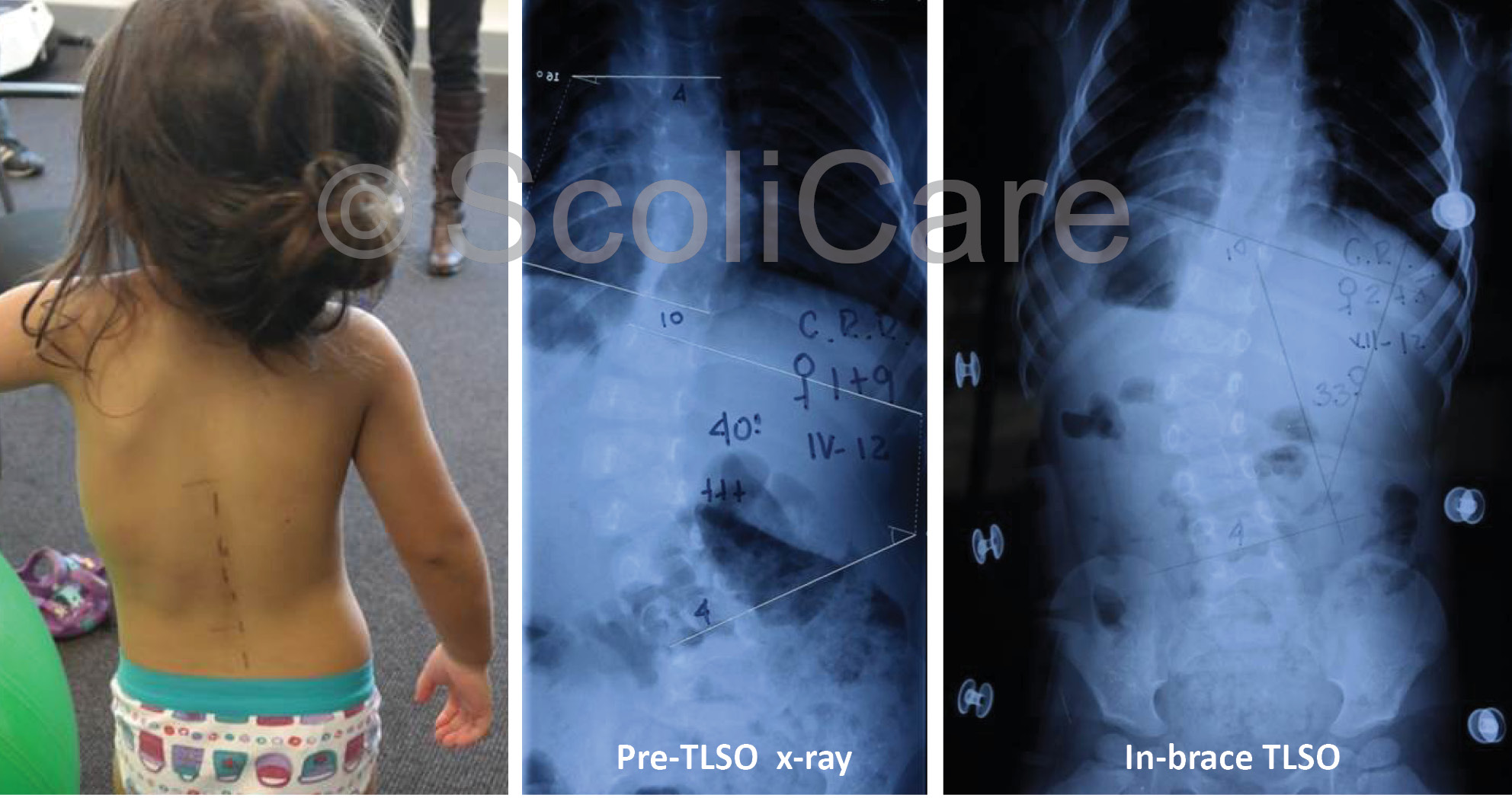
The patient presented to the ScoliCare clinic at 14 years of age with postural deformity and pain in the middle back.

Examination Findings

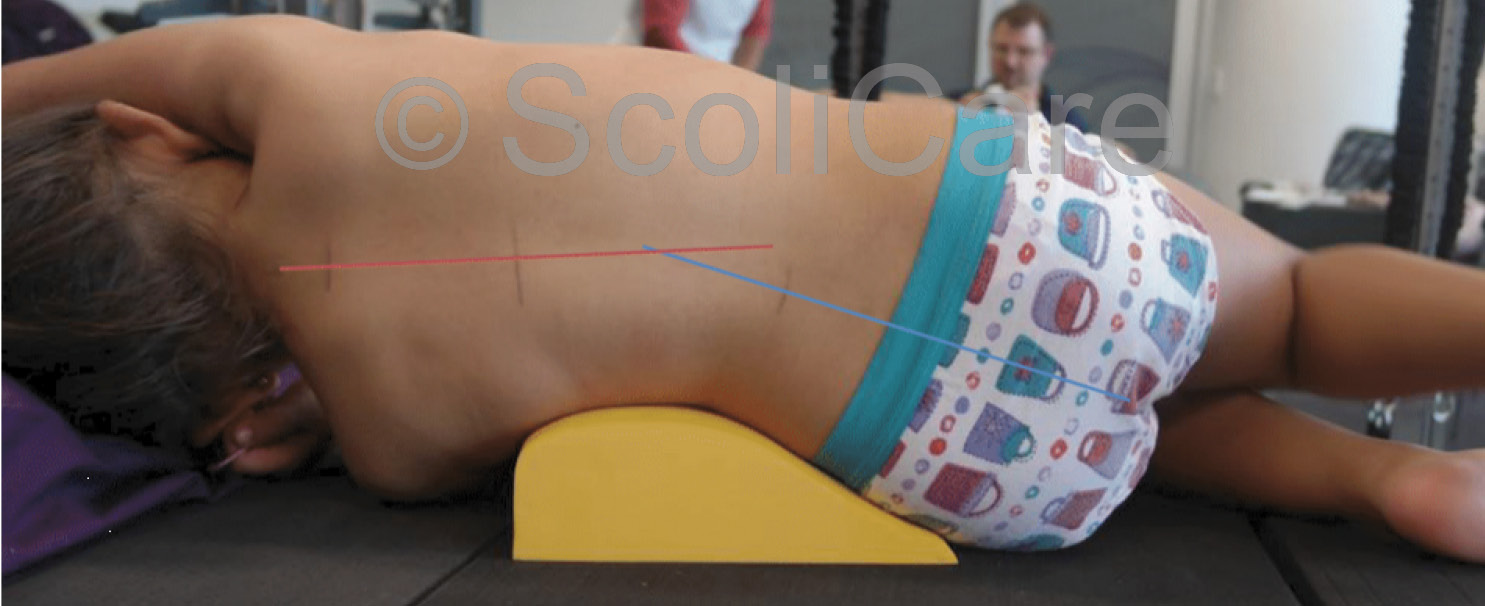
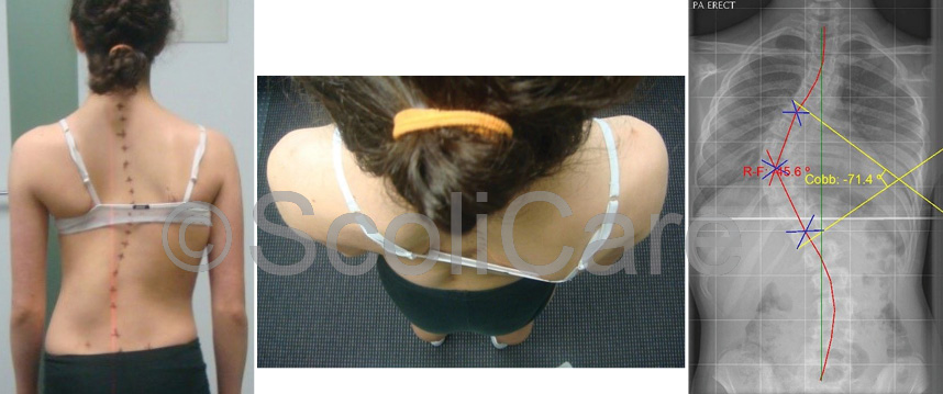
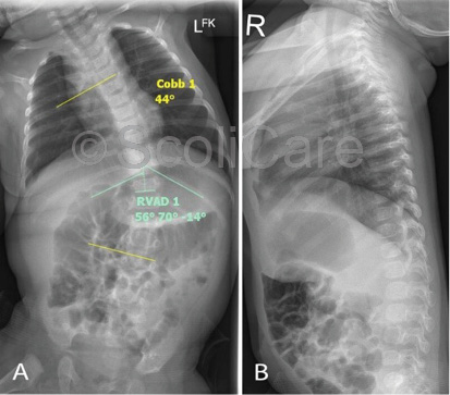
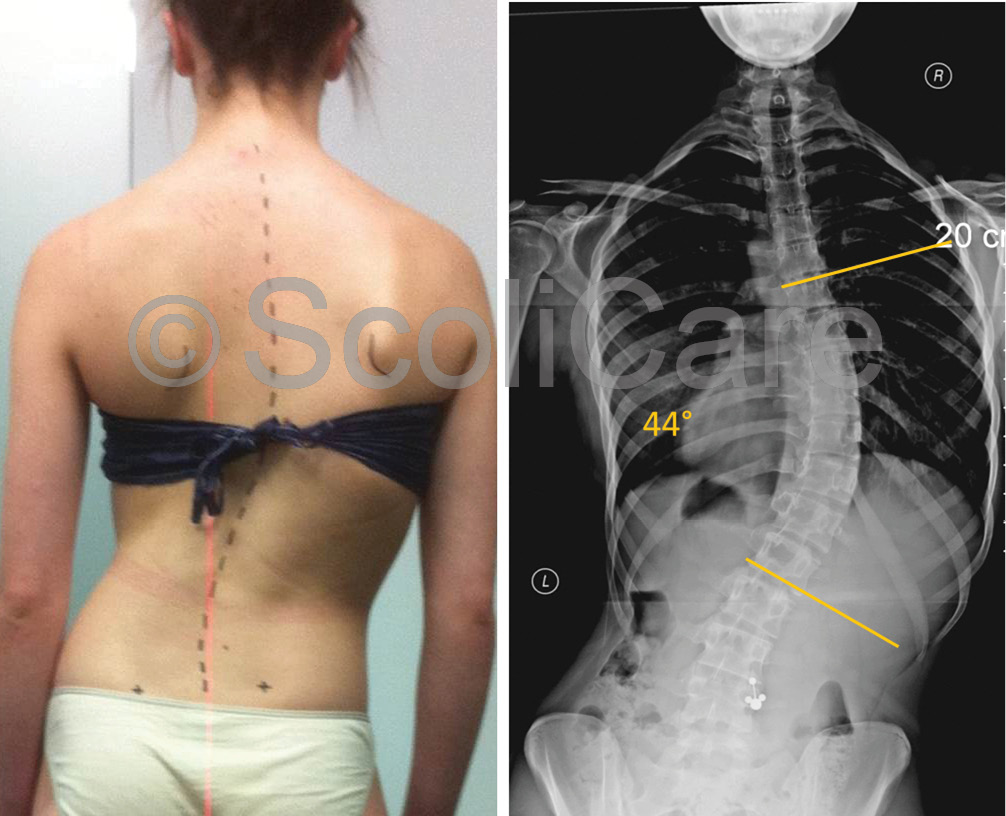
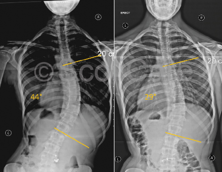
The patient was a pre-menarchal teenager who reported pain across the shoulder blades that extended from the upper thoracic spine down to the lower lumbar spine. The patient rated their pain as 6/10 on a visual analogue scale and stated that the pain and stiffness in their back was impacting on their daily activities. The patient was otherwise healthy. The physical examination revealed a significant increase in the patient’s thoracic kyphosis and lumbar lordosis (Figure 1), with marked angulation of the thoracolumbar spine during forward bending. There were no other significant clinical findings. A provisional diagnosis of Scheuermann’s kyphosis was made and confirmatory imaging was requested. Plain films taken soon after the examination highlighted an abnormal curvature in the sagittal plane – specifically a 67° hyper-kyphosis in the thoracic spine with an apex at T10. There was notable wedging of the T10 & T11 vertebrae in the lower thoracic spine.

image
Figure 1: Lateral postural photograph (Left).
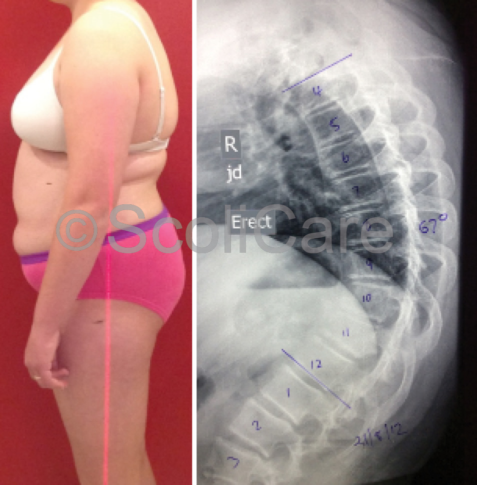
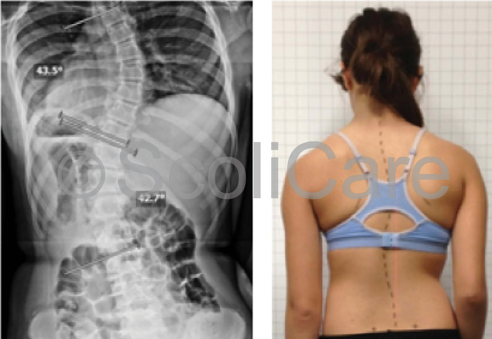
Lateral x-ray indicating a hyper-kyphosis (Right)

Intervention

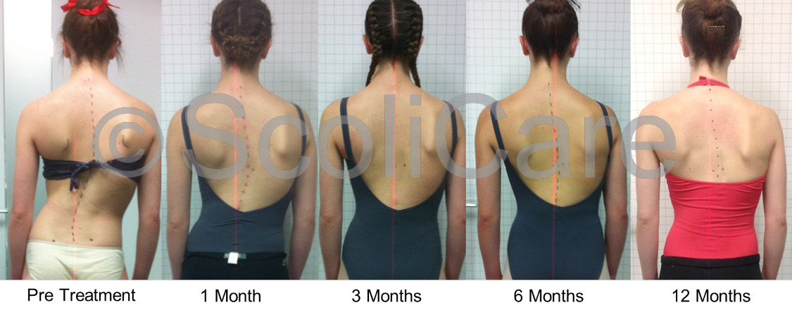
The patient was prescribed a customised 3D designed rigid kyphosis brace and specific exercise rehabilitation to address the spinal deformity. The patient was advised to wear the brace for 20 hours per day for a period of 18 months. With respect to the exercise rehabilitation, specific exercises were given to strengthen the spinal extensors. The exercises were administered during one-on-one sessions with an exercise physiologist initially, then the patient was instructed to perform these exercises daily at home to complement the action of the brace. Exercises were modified and/or progressed as the patient’s strength and endurance improved.

Outcomes

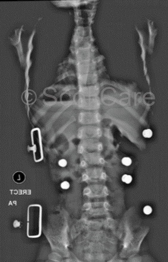
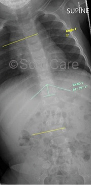
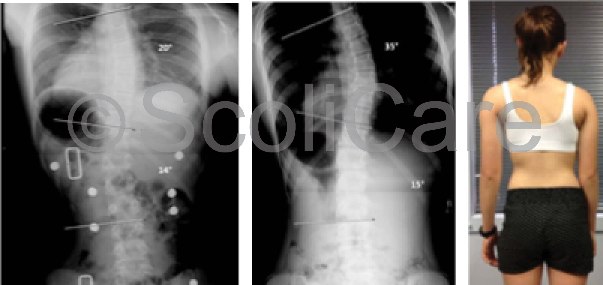
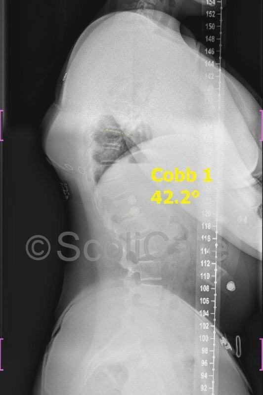
In-brace x-rays were taken one month after the brace fitting which indicated that the curve could be reduced from 67° down to 42° whilst wearing the brace (Figure 2).

After three months of brace wear an x-ray was taken with an independent radiology clinic. The images were taken with the patient standing in their normal relaxed posture after being out of the brace for 12 hours. These x-rays demonstrated a reduction in the curve from 67° down to 54°.

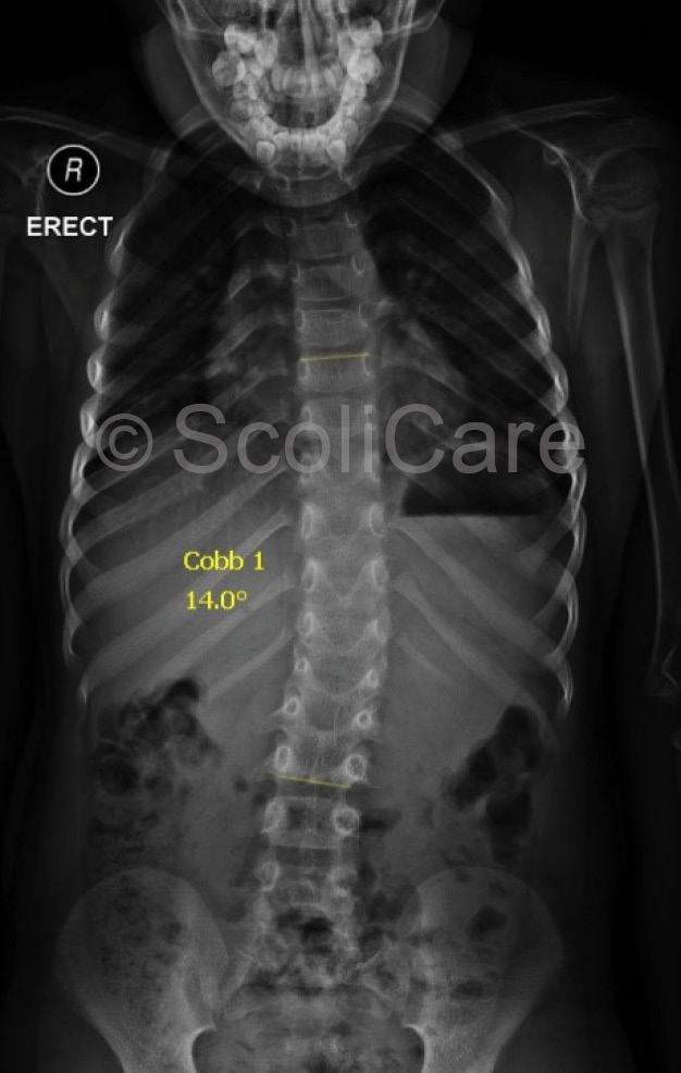
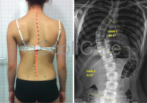
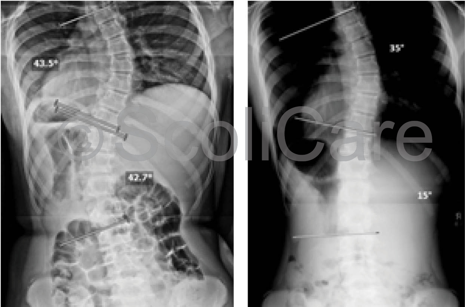
The patient wore the brace and continued the exercises for a further 12 months. Brace weaning was initiated after 15 months of brace wear. Plain films taken at the end of the brace weaning period revealed that the curve had remained stable at 54° (Figure 3). The patient was instructed to continue with the exercise rehabilitation after the cessation of brace wear, with the expectation that if the patient keeps up with their exercise program that their curve will remain under 60°.

image
Figure 2: In-brace X-rays one month


image
Figure 3: Lateral postural photograph (Left).
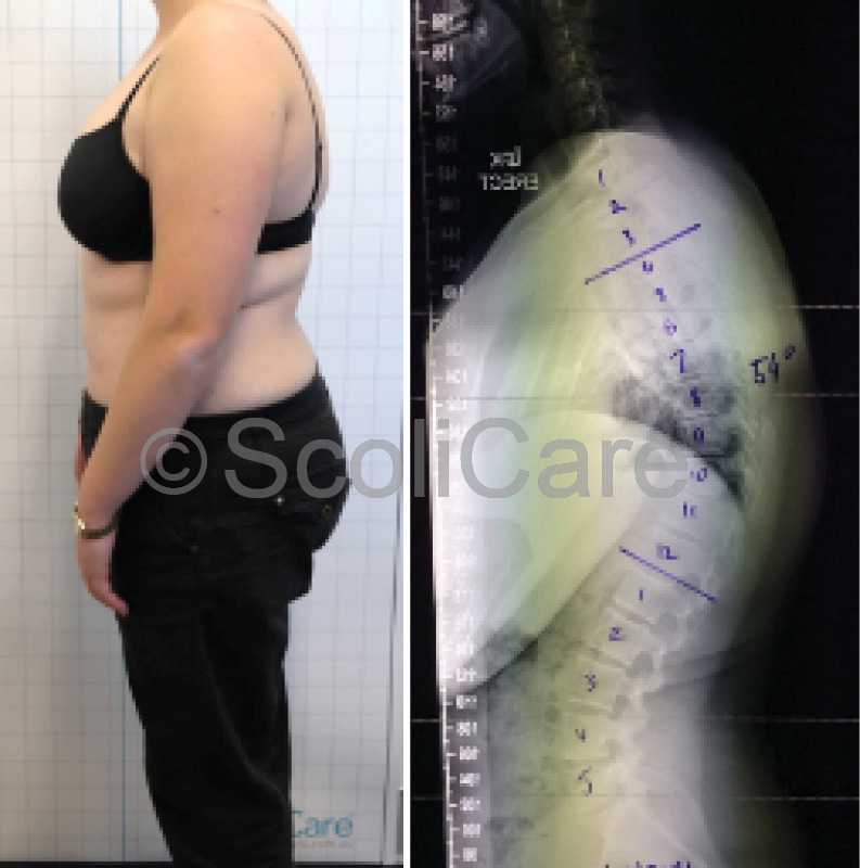
Lateral x-ray indicating 54° thoracic kyphosis  after the brace fitting 

Discussion

The normal angle of kyphosis in young patients ranges from 20-45°. One condition that may alter the normal thoracic curvature is Scheuermann’s disease. Classified as one of the osteochondroses, Scheuermann’s disease usually appears just prior to puberty with roughly equal prevalence between males and females. The condition is characterised by an increase in the normal thoracic kyphosis and is associated with structural changes in the vertebral bodies – namely wedging, endplate irregularities and diminished anterior vertebral body growth.

Long-term follow-up studies highlight that patients with Scheuermann’s disease tend to have more severe back pain, work lighter jobs and have more concerns regarding their appearance when compared with healthy patients. If left untreated, patients with kyphosis tend to experience a progression of their deformity and associated back pain. The scientific literature points to bracing being an effective treatment for patients with hyper-kyphosis. Bracing appears to not only prevent progression in this group but may also serve to restore the normal kyphosis in some patients. The best results from bracing are observed when treatment is initiated in skeletally immature patients who have curvatures of between 55-80°.

 Conclusion

This case demonstrates the successful management of an adolescent patient with hyper-kyphosis using a customised 3D designed rigid kyphosis brace and specific exercise rehabilitation.

NB: Results vary from case to case. Our commitment is to recommend the most appropriate treatment based on the patients type and severity of scoliosis.

© ScoliCare & The ScoliCare Clinic Sydney

Case – Infantile Bracing

Posted on March 19th, 2018 by admin

Treatment of an infantile scoliosis using a 3D designed scoliosis brace

Case Background

The 3-year-old female patient presented to the ScoliCare clinic with postural deformity and lateral shifting of her trunk. The deformity had been picked up by the child’s parents who had then consulted with a GP. X-ray imaging ordered by the GP revealed a left thoracolumbar curve measuring 40° and a mild (10°) compensatory right thoracic curve (Figure 1). The GP had made a diagnosis of infantile scoliosis and referred the patient on to an orthopaedic surgeon for an appraisal. After assessing the patient and reviewing the x-rays, the surgeon suggested that bracing would be the most suitable treatment in this case.

image
Figure 1: Posteroanterior postural photograph (Left), Initial posteroanterior x-ray (Middle),
In-Brace [Hospital TLSO] posteroanterior x-ray (Right)

The patient was subsequently issued with a rigid brace (thoraco-lumbo-sacral orthosis [TLSO]) that had been designed and fitted by the hospital orthotist. In-brace x-rays taken soon after the TLSO fitting showed a reduction in the size of the primary curve from 40° to 33° (Figure 1). While a reduction in the curve was evident, the parents of the patient had concerns that there was still significant deformity present despite wearing the TLSO. This prompted them to seek a second opinion from the ScoliCare clinic.

Examination Findings

The history and physical examination performed at the ScoliCare clinic aligned with the findings from the previous imaging results. The patient’s trunk was significantly translated to the left during both the postural and gait analyses, without evidence of leg length discrepancy or pelvic anomaly. The primary curve appeared flexible (Figure 2) which was a favourable sign for brace therapy.

image

Figure 2: Photograph of the patient side-lying
over a traction fulcrum orthotic device

Intervention

A new custom 3D designed scoliosis brace was prescribed for the patient along with some home based stretching and exercises to help with the abnormal trunk shift. The patient was advised to wear the brace full-time (up to 23 hours per day) and perform the rehabilitation exercises on a daily basis.

Outcomes

In-brace x-rays taken at the time of the brace fitting revealed that the deformity could be completely reduced (Figure 3). The patient continued to wear the brace with good self-reported compliance from the parents. Unfortunately, the daily exercises were performed on a more haphazard basis. Bracing treatment continued for approximately 24 months. Several modifications were made to the brace along the way to accommodate the patient’s growth.

image
Figure 3:  In-Brace (Custom 3D designed brace)
posteroanterior x-ray


By the end of the bracing period, the patient’s scoliosis had been reduced from 40° down to 14° out of the brace (Figure 4).At this point in time the patient is still under care, however the recommendation is for the patient to wear a more flexible style brace to maintain the correction achieved with the custom 3D designed brace, and also to further reduce the left postural shift.

image
Figure 4: Out-of-brace x-ray

 Discussion

This case highlights the management of an infantile idiopathic scoliosis (IIS) case using a custom 3D designed scoliosis orthosis. Patients with IIS typically present before the age of 3 years and are classified into two categories – resolving and progressive. Curves in patients with the resolving type tend to reduce spontaneously over time, whereas curves in patients with the progressive type continue to worsen leading to crippling deformity and reduced quality of life if not treated. Casting is typically initiated before the age of two years in these patients as it is more difficult to attain a complete resolution of the deformity if treatment is initiated after this point. After a significant result has been obtained with casting, the patient is usually placed into a rigid brace to stabilise the correction.

There are unique features to this particular case. Thoracolumbar presentations are less common compared to thoracic presentations in IIS patients, and the presence of compensatory curves is also somewhat unusual in this population. In this case, bracing has been used as a first-line management approach versus the more traditional approach involving serial casting. The patient’s initial hospital-made-brace was substandard as evidenced by the small (17.5% reduction) in-brace correction, and had the patient continued with this brace it is likely that the curve would have continued to progress.

At this point in time the evidence to support the use of bracing as a primary treatment in patients with IIS is sparse, however the results observed in this case would suggest that a more comprehensive investigation of ‘over corrective’ 3D bracing is justified.

Conclusion

This case demonstrates the reduction of a severe thoracolumbar scoliosis in a young child using a custom 3D designed scoliosis orthosis.

NB: Results vary from case to case. Our commitment is to recommend the most appropriate treatment based on the patients type and severity of scoliosis.

© ScoliCare

Case – Bracing & Exercise Rehab

Posted on February 16th, 2018 by admin

Reduction of a severe scoliosis in a young female gymnast using a 3D designed scoliosis brace

Case Background

The patient was a 14-year-old, high-level, competitive gymnast. The patient’s mother had noticed a curvature in her daughter’s spine and taken her to see a manual therapist. The therapist had made a diagnosis of Adolescent Idiopathic Scoliosis (AIS) and treated the patient for three months. Unfortunately, the patient had not responded to treatment and was referred to the ScoliCare clinic for an assessment.

Examination Findings

The examination revealed some significant postural changes. Specifically, notable curvature in the middle and lower sections of the patient’s spine, uneven heights of the pelvic bones, and torsion of the rib cage (Figure 1). X-rays taken at the time showed a large 71° curve in the thoracic spine and a secondary curve, measuring 40°, in the lumbar spine (Figure 1). The patient was only Risser 2, indicating that she still had a significant amount of growing to do.

image
Figure 1: A) Posterior-anterior view highlighting the scoliosis and pelvic tilt.
B) Superior to inferior view highlighting torsion of the rib cage associated with the thoracic spine curvature.
C) Posterior-anterior x-ray demonstrating the curvature of the spine and changes in the rib cage.
  

Intervention

As both the curves were quite large, and the patient was skeletally immature, it was recommended that the patient be seen by an orthopaedic surgeon. The surgeon’s recommendation was that the patient undergo surgery to correct the abnormal spinal curvatures. After careful consideration, both the patient and her parents declined the surgeon’s recommendation. Instead, they decided to go with a more conservative approach.

The patient was subsequently prescribed a customised 3D designed rigid brace , and rehabilitation exercises based on the SCHROTH principles. The recommendation was that the patient be braced for two years until she stopped growing. A daily scoliosis specific exercise program was designed for the patient to support the corrections being made by the brace. New exercises were added to the program as the patient progressed. Regular follow-ups were scheduled to ensure that A) the patient was responding to the treatment, and B) to monitor compliance with the bracing and exercise program.

Outcomes

The patient’s spine responded favourably to the treatment with one-month x-rays showing that the larger curve could be reduced from 71° down to 40° with the patient wearing the brace (Figure 2).

In total, the patient was braced for 18 months. The patient also participated in supervised exercise sessions at the clinic and performed similar exercises at home. At the end of the treatment period, the patient’s curves had reduced significantly (71° to 56°) as demonstrated in the out-of-brace x-rays in Figure 3. The patient’s posture was also notably improved.

image

Figure 2: In-brace frontal x-rays taken one
month after the brace fitting.

image

 Figure 3: A) Post-treatment posterior-anterior view posture photo B) Post-treatment frontal x-rays.

Discussion

Normally, brace treatment is reserved for AIS patients with moderate (20-45°) scoliotic curves. Scoliosis specific exercise rehabilitation is used in mild (10-20°) scoliosis as a standalone treatment, and as a supplement to bracing in patients with moderate scoliosis. Patients with more severe scoliosis are typically managed surgically. There is very little research to support the use of bracing or exercise in patients with curves exceeding 45°. This case is unique in the sense that the patient/parents refused surgery and elected to try a more conservative option. Care was taken to explain to the patient/parents that surgical treatment is the recommended pathway for AIS patients with severe curves. The is a scarcity of evidence to support the use of bracing in curves >60° in this population.

Conclusion

This case demonstrates the reduction of a severe scoliosis in an AIS patient who declined surgical intervention.

NB: Results vary from case to case. Our commitment is to recommend the most appropriate treatment based on the patients type and severity of scoliosis.

© ScoliCare & The ScoliCare Clinic Sydney


Case – Infantile Bracing

Posted on February 16th, 2018 by admin

Resolution of a moderate scoliosis in an infant patient using a 3D designed scoliosis brace

Case Background:

The patient presented to the ScoliCare clinic at 11 weeks of age with a marked ‘C’ shaped scoliosis. The patient’s mother had noticed the curvature in her daughter’s spine soon after birth. The child had been delivered naturally, however the child had “gotten stuck” and support staff had needed to intervene. The child’s shoulder was dislocated at this time, and later relocated. The mother had taken her daughter to the local chiropractor approximately 1.5 months after the birth for an assessment. The chiropractor had provided a short course of treatment and prescribed gentle bending exercises to help reduce the scoliosis. Unfortunately, the scoliosis had not responded to treatment and was referred to the ScoliCare clinic.

Examination Findings

The examination performed at the ScoliCare clinic revealed a single, large, left ‘C’ curve (Figures 1A & B). The patient also had difficulty turning their head to the right. The curve was very stiff with little correction during side-bending or traction. The neurological exam was normal except for a slight reduction in the abdominal reflexes. X-rays ordered soon after confirmed the findings from the physical exam. The single, left, thoracolumbar curve measured 44° Cobb with an apex at T11. The patient was diagnosed with infantile idiopathic scoliosis and was referred to an orthopaedic surgeon for an evaluation.

image
 Figure 1: Postural photographs of the infant at the time of the initial consultation


image
Figure 2: Frontal and lateral x-rays

Intervention

There was a long wait before the patient could be seen by the surgeon, and as a result the parents decided to start with conservative treatment.

A customised 3D designed rigid brace was prescribed and the patient was advised to continue with the exercises that had been advised by the chiropractor. The patient was instructed to wear the brace for short periods of time at first and then build up the time wearing the brace up to a maximum of 8 hours.

The parents were advised that the brace was only to be worn under supervision, and only during the day. Eventually, the infant was seen by the orthopaedic surgeon who recommended physiotherapy. The parents however chose to continue with the bracing and exercise program.

Outcomes

The patient wore the custom scoliosis brace on a part-time schedule for eight months. At the end of the bracing period, the child’s scoliosis had been reduced from 44° down to 7° (Figure 4).

A follow-up examination was performed 23 months after the initial presentation. The angle of trunk rotation measured 0° at this time and there was no evidence of scoliosis or movement abnormalities.

The parents were very happy with the result and reported that, although challenging at times, the treatment was tolerated by the child.

image
Figure 4: Frontal x-ray taken after the patient
had been out 
of the brace for four months

Discussion:

Infantile idiopathic scoliosis presents before the age of 3 years and is classified into two types – resolving and progressive. Curves in patients with the resolving type tend to reduce spontaneously over time, whereas curves in patients with the progressive type continue to worsen leading to crippling deformity and reduced quality of life if not treated. There are x-ray markers that can be used to differentiate between the two types. However, the difficult aspect for clinicians dealing with patients with idiopathic infantile scoliosis is that the two types can present identically in the initial stages.

Observation is recommended as the initial action, however this can be challenging for parents and clinicians as they wait for a worsening in the patient’s deformity, which, may already be quite significant. For patients with the progressive type of infantile scoliosis, the standard treatment involves placing the infant in a straightened position then applying a plaster cast.

As the child grows, the casts are removed and replaced. This process is repeated, for years in some cases, until the scoliosis has been reduced. The casting process involves intubating and placing the infant under a general anaesthetic. A surgeon, anaesthetist, nursing staff and a specialist table are required for the procedure, which takes approximately 90 minutes. While casting is successful in the management of most cases of infantile idiopathic scoliosis, there are concerns that the frequent exposure to anaesthetising agents may be damaging to the developing brain of the infant. Treatments such as bracing may represent a safer alternative, but further research is required in this area.

Conclusion:

This case study demonstrates the reduction of a moderate infantile idiopathic scoliosis using a customised 3D designed scoliosis brace.

NB: Results vary from case to case. Our commitment is to recommend the most appropriate treatment based on the patients type and severity of scoliosis.

© ScoliCare & The ScoliCare Clinic Sydney


Case – Bracing and Exercise Rehabilitation

Posted on January 27th, 2017 by admin

Patient aged 13 years with a 33 degree left thoracolumbar scoliosis.

Background:

This patient was 13 years old at the time of diagnosis. She had noticed her posture was off centre and was experiencing mild back pain. Sports participation included ballet, other dance styles and netball. The Scoliosis was diagnosed 4-5 weeks before the initial consultation at Sydney Scoliosis Clinic.

The medical history was unremarkable with the neurological examination within normal limits. Birth and developmental milestones were within normal limits (as reported by her mum). There was tenderness to palpation of the spine at the Thoracolumbar junction. There was pain on left lateral flexion of the spine.

The patients’ grandfather was diagnosed with Scoliosis.

Initial X-rays showed a 33 degree left thoracolumbar scoliosis. There was also significant lumbar spine rotation on x-ray. The Risser sign on the pelvis was Risser 1-2, indicating that the patient was skeletally immature and more growth and progression was expected without treatment.

image

Treatment:

As the patient was still growing, was a Risser 1 and the curve was greater than 30 degrees, there was a high risk of progression. A combination of a customised 3D designed rigid brace, scoliosis specific rehabilitation exercises and use of a scoliosis orthotic device were recommended.

The scoliosis brace was to be worn full-time. This is 23 hours per day with extra time up to a maximum of 4 hours out of the brace if the patient was actively participating in sports during those out of brace hours. The weaning into the brace was started with 2 hours of brace wear on the first day followed by adding another 2 hours every subsequent day until the required full-time hours were attained.

The scoliosis specific exercises were initially implemented as twice a week for 3 weeks, followed by once per month. The patient was required to complete the exercise protocol at home daily.

A scoliosis orthotic device was also used daily for 20 minutes to stretch the spine at the Thoracolumbar junction. This device was placed at the left apex with the patient lying on her left side. Exercises were performed on the device.

Results:

At the one month in-brace x-ray the curve had reduced to 13 degrees. At the 3 month out of brace x-ray the curve had reduced to 26 degrees. At this point extra corrective padding was added to the brace to increase the 3-dimensional corrective action of the brace. At the 12 month mark an out-of-brace x-ray was taken. The results of which showed that the spine was 11 degrees without using the brace.

The last x-ray was taken 22 months after the start of treatment. This x-ray was an out-of-brace x-ray where the patient was required to be out of the brace for at least 6 hours. The results of which show her spine to have a 6 degree curvature, which according to definition cannot be classified as a scoliosis.

image

The postural assessment showed continuous improvements of her posture with her body showing good balance after 4 months of treatment to the point that she was symmetrical by the 12 month mark. The postural improvements have been maintained throughout the treatment period.

The functional assessment of fatigue ability and strength of her core muscles, together with the flexibility of her spine showed no deterioration of strength, endurance or flexibility.

Conclusion:

A rigid custom designed 3D scoliosis brace, in combination with a  scoliosis specific rehabilitation program, may achieve reduction in Cobb angle in similar cases.  This approach follows the SOSORT criteria as was shown in the literature review to have a positive prognostic impact on Thoracolumbar curvatures.

This approach is a team approach that allows the patient to learn a scoliosis specific exercise program, together with a bracing technique that does not use a 3 point pressure system, but rather is an over-corrective custom designed brace that is designed to address all aspects of the scoliosis from sagittal and coronal balance to lateral translation at the apex and vertebral rotation of the curve.

This patient is still undergoing treatment and is expected to wear their scoliosis brace a minimum of 20 hours per day for 2 years in total. The prognosis for this patient is to maintain a similar amount of correction as seen at the 22 month mark.

NB: All cases are different and results may vary case to case. Our commitment is to recommend the most appropriate treatment based on the patient’s type and severity of scoliosis.

© ScoliCare & Sydney Scoliosis Clinic 2016


Case – Bracing and Exercise Rehabilitation

Posted on September 11th, 2015 by admin

Patient aged 17 with scoliosis curve measured 44 degrees reduced to 29 degrees.

Background: 

Patient presented to the Sydney Scoliosis Clinic aged 17 at the time of initial examination. Adolescent Idiopathic Scoliosis was diagnosed 18 months earlier and no treatment was undertaken during this time. At the initial consultation the  scoliosis curve measured 44 degrees. This patient had seen a surgeon and was recommended to undergo surgery.  She reported having bad back pain (7-8/10) most days. The pain was worse after dance classes.

The patient was on track to become a professional ballet dancer however the pain and the effect of the scoliosis on the body posture and the effect that surgery would have on the spinal flexibility meant that this future career was at risk.

image

Treatment:

A combination of customised 3D designed rigid brace wear, and a scoliosis specific exercise program based on the SEAS principles was recommended.  It was recommended to use the brace for 6-8 hours per day and to do the exercises twice per day for 20 minutes. This intensive regime was recommended for 3-6 months.

Results:

As seem from x-rays and posture photos, a cosmetic improvement in the body was seen within the first 4 weeks.  This improvement was maximised at 6 months. After the end of intensive treatment, the body posture settled to its new corrected position which is demonstrated at the 12 months photo.  At the 12 month mark an x-ray was taken with an independent radiology clinic with the patient standing in her relaxed normal posture.  This shows an improvement in the scoliosis curve and the alignment of the spine.  The curve was corrected by 15 degrees, reducing from 44 degrees to 29 degrees.

The patient’s pain was reduced to 2-3/10 and was only present on an occasional basis. As the body posture improved so did the performance at ballet. At the end of the intensive part of the treatment program the patient was selected to join a top ballet institution overseas.  As a result of these improvements surgery was cancelled.

Discussion:

This case demonstrates the potential for a combination of customised 3D designed rigid brace wear and scoliosis specific exercise to improve the cosmetic look of the body, the spinal alignment and degree of curve, and the pain associated with the scoliosis. Although relatively mature this patient was able to achieve 15 degrees of correction of her curve.

Although this may not be achievable in every case it shows that in cases where the patient is flexible and there is an aspect of the scoliosis that is not fixed it may be possible to make correction even after growth has finished. Continuation of the exercise program may be required to maintain the correction.

image

Conclusion:

In this case it was possible to reduce a large curve that was at a surgical level to a moderate curve no longer in need of surgery. Choosing the right treatment to achieve the goals of cosmetic improvement, curve reduction, and pain relief was successful in this case.

image


NB: All cases are different and results may vary case to case. Our commitment is to recommend the most appropriate treatment based on the patient’s type and severity of scoliosis.

© ScoliCare 2015

Case – Bracing and Exercise Rehabilitation

Posted on September 10th, 2015 by admin

Patient aged 13 with scoliosis curve measured 44° in the thoracic spine and 43° in the lumbar spine reduced to 35° and 15° in 8 months!

Background:

Patient presented to the Sydney Scoliosis Clinic aged 13 at the time of initial examination. Postural issues had been noticed 6 months earlier. At the initial consultation the scoliosis curve measured 44° in the thoracic spine and 43° in the lumbar spine. Her pelvic growth plates were open and graded as Risser stage 1. Adolescent Idiopathic Scoliosis was diagnosed. This patient had been recommended to undergo surgery.

image

Treatment:

A combination of full time bracing using a customised 3D designed rigid brace and a daily home rehabilitation program, using a scoliosis treatment aid and Mirror Image exercises, was recommended.  It was recommended to wear the brace for 23 hours per day and to use the treatment aid once per day for 20 minutes. This treatment regime was recommended for a minimum 24 months as the patient was immature and had substantial growth remaining.

Results:

As can be seen from the photos, a cosmetic improvement in the body was seen within the first 3 months. This improvement was maintained at 12 and 18 months. At the 3 month mark an in-brace x-ray was taken showing very good correction of the curve. The thoracic curve reduced from 44 to 20 degrees and the lumbar curve reduced from 43 to 14 degrees (in the brace).

image


At the 8 month mark an out-of-brace x-ray was taken at an independent radiology clinic, with the patient standing in her relaxed normal posture. This showed a significant improvement in the scoliosis curves. The lumbar curve was corrected by a staggering 28 degrees!

The thoracic spine curve was corrected by 9°.

The patient has continued with compliant treatment, and postural photographs at the 12 and 18 month mark show stability of the result. She is continuing with treatment.

Discussion:

Xrays and posture photos from this case have shown that this combination of treatment has been able to improve the cosmetic look of the body, the spinal alignment and the degree of curve. Although still immature and undergoing treatment, this case represents a positive initial result that will likely result in this patient avoiding spinal fusion surgery in the future.

Conclusion:

A rigid custom designed 3D scoliosis brace, in combination with a scoliosis specific exercise program, may achieve reduction in Cobb angle in similar cases. Choosing the right treatment with the aim of curve reduction and cosmetic improvement, are so far proving to be successful in this case.

image


NB: Results vary from case to case. Our commitment is to recommend the most appropriate treatment based on the patient’s type and severity of scoliosis.

© ScoliCare 2015