Posts Tagged "ScoliBrace"

Case – Bracing and Exercise Rehabilitation

Posted on January 27th, 2017 by admin

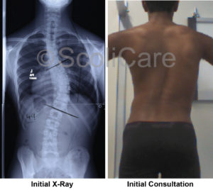
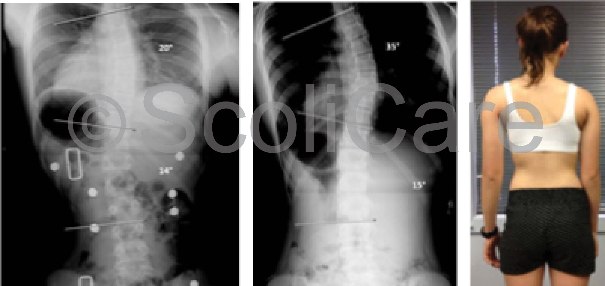
Patient aged 13 years with a 33 degree left thoracolumbar scoliosis.

Background:

This patient was 13 years old at the time of diagnosis. She had noticed her posture was off centre and was experiencing mild back pain. Sports participation included ballet, other dance styles and netball. The Scoliosis was diagnosed 4-5 weeks before the initial consultation at Sydney Scoliosis Clinic.

The medical history was unremarkable with the neurological examination within normal limits. Birth and developmental milestones were within normal limits (as reported by her mum). There was tenderness to palpation of the spine at the Thoracolumbar junction. There was pain on left lateral flexion of the spine.

The patients’ grandfather was diagnosed with Scoliosis.

Initial X-rays showed a 33 degree left thoracolumbar scoliosis. There was also significant lumbar spine rotation on x-ray. The Risser sign on the pelvis was Risser 1-2, indicating that the patient was skeletally immature and more growth and progression was expected without treatment.

image

Treatment:

As the patient was still growing, was a Risser 1 and the curve was greater than 30 degrees, there was a high risk of progression. A combination of a customised 3D designed rigid brace, scoliosis specific rehabilitation exercises and use of a scoliosis orthotic device were recommended.

The scoliosis brace was to be worn full-time. This is 23 hours per day with extra time up to a maximum of 4 hours out of the brace if the patient was actively participating in sports during those out of brace hours. The weaning into the brace was started with 2 hours of brace wear on the first day followed by adding another 2 hours every subsequent day until the required full-time hours were attained.

The scoliosis specific exercises were initially implemented as twice a week for 3 weeks, followed by once per month. The patient was required to complete the exercise protocol at home daily.

A scoliosis orthotic device was also used daily for 20 minutes to stretch the spine at the Thoracolumbar junction. This device was placed at the left apex with the patient lying on her left side. Exercises were performed on the device.

Results:

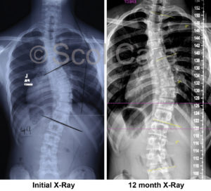
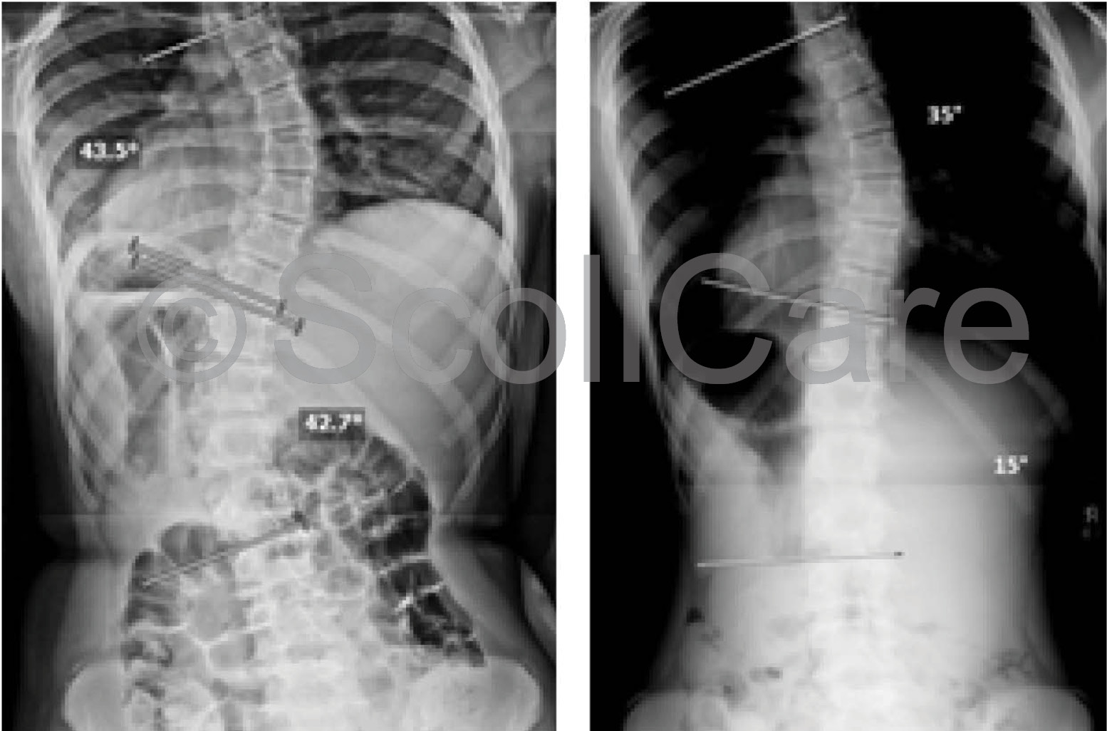
At the one month in-brace x-ray the curve had reduced to 13 degrees. At the 3 month out of brace x-ray the curve had reduced to 26 degrees. At this point extra corrective padding was added to the brace to increase the 3-dimensional corrective action of the brace. At the 12 month mark an out-of-brace x-ray was taken. The results of which showed that the spine was 11 degrees without using the brace.

The last x-ray was taken 22 months after the start of treatment. This x-ray was an out-of-brace x-ray where the patient was required to be out of the brace for at least 6 hours. The results of which show her spine to have a 6 degree curvature, which according to definition cannot be classified as a scoliosis.

image

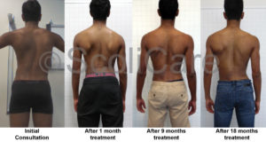
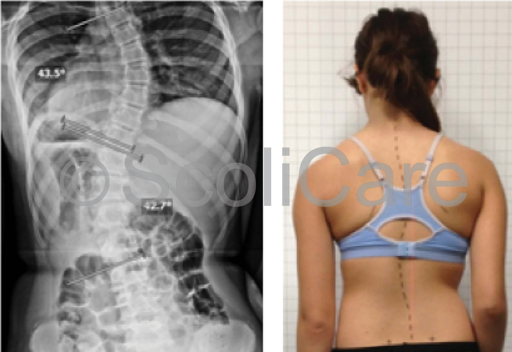
The postural assessment showed continuous improvements of her posture with her body showing good balance after 4 months of treatment to the point that she was symmetrical by the 12 month mark. The postural improvements have been maintained throughout the treatment period.

The functional assessment of fatigue ability and strength of her core muscles, together with the flexibility of her spine showed no deterioration of strength, endurance or flexibility.

Conclusion:

A rigid custom designed 3D scoliosis brace, in combination with a  scoliosis specific rehabilitation program, may achieve reduction in Cobb angle in similar cases.  This approach follows the SOSORT criteria as was shown in the literature review to have a positive prognostic impact on Thoracolumbar curvatures.

This approach is a team approach that allows the patient to learn a scoliosis specific exercise program, together with a bracing technique that does not use a 3 point pressure system, but rather is an over-corrective custom designed brace that is designed to address all aspects of the scoliosis from sagittal and coronal balance to lateral translation at the apex and vertebral rotation of the curve.

This patient is still undergoing treatment and is expected to wear their scoliosis brace a minimum of 20 hours per day for 2 years in total. The prognosis for this patient is to maintain a similar amount of correction as seen at the 22 month mark.

NB: All cases are different and results may vary case to case. Our commitment is to recommend the most appropriate treatment based on the patient’s type and severity of scoliosis.

© ScoliCare & Sydney Scoliosis Clinic 2016


Case – Bracing and Exercise Rehabilitation

Posted on October 23rd, 2015 by admin

Male patient aged 14 years with a right thoracic curve measured at 49 degrees.

Background 

This patient was diagnosed with Scoliosis by his Physiotherapist at the age of 13 years old with a curve of 39 degrees. They first consulted a surgeon about the case who suggested surgery or bracing.

A year later they presented to the Sydney Scoliosis Clinic for bracing and the curve had progressed a further 10 degrees. The patient was not experiencing any pain from the scoliosis, but did notice the asymmetrical shape of his torso. This patient is a National medallist in Athletics and also previously played representative Australian Rules Football. Due to his regular training, this patient had a good general strength and flexibility.

1- Background

Treatment

This patient’s treatment involved wearing a customised rigid brace for 23 hrs per day, ongoing scoliosis specific exercise sessions and tractioning exercises with a scoliosis treatment aid. His brace wear started at 2 hrs per day, which was built up gradually over the following weeks to 23 hours each day. The patient does not compete in the brace but is very diligent in putting it back on straight after an event.

The scoliosis specific exercises require him to position his body in an active self-correction, which is the mirror image of his scoliosis, and to also work on strengthening his core and lower back muscles with exercises specifically prescribed for his case. He regularly sees the exercise physiologist to assess his progress and increase the difficulty of the exercises to ensure he keeps improving.

The patient does quite advanced  exercises and we have now started incorporating his active self-correction to warm-up movements he does at training to help keep his spine in good alignment when he is out of the brace. The home exercises are done every day and take approximately 40mins to complete.

Results

From examining xrays and posture photos, there has been an improvement in this patient’s posture in the first 12 months of treatment. The intial curve of 49 degrees, has now decreased to 35 degrees.

3a - Results

Due to his age and currently being Risser 4, it is essential for the brace treatment to continue while he is still growing. If left untreated, the scoliosis can progress further with growth.

At the beginning of the treatment, the patient was not competing at his best, but after a period of adaptation and the inclusion of specific exercises, he is now excelling and is back to the optimal form he was in prior to starting his scoliosis treatment.

The patient and his family are very committed to his treatment and to helping him succeed.

3b - Results


Conclusion

This  case demonstrates the reduction of a large curve that was at a surgical level to a moderate curve no longer at risk of needing surgery. The combination of a rigid custom designed 3D scoliosis brace, with scoliosis specific exercise, may achieve reduction in curve and cosmetic improvement in similar cases.

The patient was also able to continue competing at an elite level without compromising his ability.

NB: All cases are different and results may vary case to case. Our commitment is to recommend the most appropriate treatment based on the patient’s type and severity of scoliosis.

© ScoliCare 2015

Case – Bracing and Exercise Rehabilitation

Posted on September 10th, 2015 by admin

Patient aged 13 with scoliosis curve measured 44° in the thoracic spine and 43° in the lumbar spine reduced to 35° and 15° in 8 months!

Background:

Patient presented to the Sydney Scoliosis Clinic aged 13 at the time of initial examination. Postural issues had been noticed 6 months earlier. At the initial consultation the scoliosis curve measured 44° in the thoracic spine and 43° in the lumbar spine. Her pelvic growth plates were open and graded as Risser stage 1. Adolescent Idiopathic Scoliosis was diagnosed. This patient had been recommended to undergo surgery.

image

Treatment:

A combination of full time bracing using a customised 3D designed rigid brace and a daily home rehabilitation program, using a scoliosis treatment aid and Mirror Image exercises, was recommended.  It was recommended to wear the brace for 23 hours per day and to use the treatment aid once per day for 20 minutes. This treatment regime was recommended for a minimum 24 months as the patient was immature and had substantial growth remaining.

Results:

As can be seen from the photos, a cosmetic improvement in the body was seen within the first 3 months. This improvement was maintained at 12 and 18 months. At the 3 month mark an in-brace x-ray was taken showing very good correction of the curve. The thoracic curve reduced from 44 to 20 degrees and the lumbar curve reduced from 43 to 14 degrees (in the brace).

image


At the 8 month mark an out-of-brace x-ray was taken at an independent radiology clinic, with the patient standing in her relaxed normal posture. This showed a significant improvement in the scoliosis curves. The lumbar curve was corrected by a staggering 28 degrees!

The thoracic spine curve was corrected by 9°.

The patient has continued with compliant treatment, and postural photographs at the 12 and 18 month mark show stability of the result. She is continuing with treatment.

Discussion:

Xrays and posture photos from this case have shown that this combination of treatment has been able to improve the cosmetic look of the body, the spinal alignment and the degree of curve. Although still immature and undergoing treatment, this case represents a positive initial result that will likely result in this patient avoiding spinal fusion surgery in the future.

Conclusion:

A rigid custom designed 3D scoliosis brace, in combination with a scoliosis specific exercise program, may achieve reduction in Cobb angle in similar cases. Choosing the right treatment with the aim of curve reduction and cosmetic improvement, are so far proving to be successful in this case.

image


NB: Results vary from case to case. Our commitment is to recommend the most appropriate treatment based on the patient’s type and severity of scoliosis.

© ScoliCare 2015